CHOLESTEATOMA
Definition: Cholesteatoma is the presence of squamous epithelial pocket or sac, filled with keratin debris within the middle ear cleft. It is a cause of Active Squamous epithelial type of Chronic otitis media.
Origin of Cholesteatoma:
- Presence of congenital cell rests.
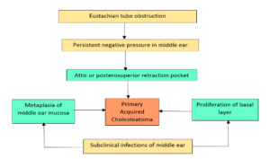
- Invagination of tympanic membrane in the form of retraction pockets (Wittmaack’s theory).
- Basal cell hyperplasia (Ruedi’s theory)– The basal cells grows under the influence of infection and forms keratinizing squamous epithelium.
- Epithelial invasion (Habermann’s theory)– The epithelium grows inside middle ear through a pre-existing perforation especially of marginal type.
- Metaplasia (Sade’s theory)– Middle ear respiratory mucosa undergoes metaplasia due to repeated infections and changes into squamous epithelium.
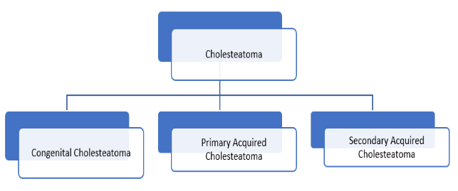
Cholesteatoma is divided into 3 types:

Congenital Cholesteatoma: They are squamous epithelial cells arising from temporal bone. It arises from ectodermal cell nest.
It is classified into:

Clinical features:
Patient presents with:
- Severe sensorineural hearing loss.
- Facial weakness.
- X-ray mastoid Schuller’s view shows bone erosion.
- HRCT scan plain 1mm cuts will be noting the extent of disease and bony erosion in temporal bone.
- MRI temporal bone plain and contrast with facial nerve studies are useful in advanced cases.
Acquired Cholesteatoma:
- Primary:
- Invagination of Pars flaccida.

- Basal cell hyperplasia.
- Squamous metaplasia.

- Invagination of Pars flaccida.
- Secondary:
- Migration of squamous epithelium from external auditory canal and tympanic membrane through the perforation present in the tympanic membrane.
- Metaplasia due to repeated infections of middle ear through the pre-existing tympanic membrane perforation.

Clinical features:
- Otorrhoea: The discharge is scanty, foul smelling, continuous and yellowish.
- Deafness: There is moderate to severe hearing loss. It may be conductive and mixed.
- Otalgia.
- Tinnitus.
- Vertigo.
- Patients may also present with intracranial, or extracranial complications.

Investigation:
- Routine blood count, X-ray chest, ECG.
- Suction microscopy: to clear out any discharge, confirm clinical findings and examination of the discharge for pus culture and antibiotic sensitivity.
- Tuning fork tests: They help in clinical evaluation of hearing loss.
- Pure tone audiometry: this is useful for a qualitative and quantitative analysis of the patients hearing. It also helps for pre-operative and post-operative assessment of the hearing loss and for medico-legal purposes.
- X-ray mastoid Schuller’s view: to diagnose the extent of the disease and status of the mastoid air cells, sinus plate, dural plate and sinodural angle.
- HRCT temporal bone is extremely useful in patients with complications or impending complications.
- MRI temporal bone plain with contrast helps diagnosing soft tissue extension.
Cholesteatoma Treatment
Surgery is the only treatment for Cholesteatoma. Medical line of treatment may be only useful to control rapid spread of infection and impending intracranial complications due to infection. Mastoidectomy: Depending on the extent and severity of the discharge, a Modified Radical or Radical mastoidectomy is indicated.
Aims of surgery:
- Make the ear safe and prevent complications.
- Make the ear dry.
- Restore maximum possible hearing.
We Are Always Ready to Help You.
Book An Appointment

