NASOPHARYNGEAL ANGIOFIBROMA
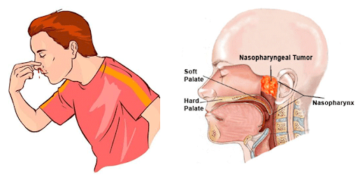
Definition: They are benign, very vascular and biologically aggressive tumors, originating almost exclusively from posterior nasal and nasopharyngeal region in adolescent males.
Aetiology:
Age– Usually occurs in the 2nd decade. Sex– Exclusively common in males. Theories of origin:
Age– Usually occurs in the 2nd decade. Sex– Exclusively common in males. Theories of origin:
- Fibroblastic Theory: This theory is suggestive of an abnormal growth or response of connective tissues such as embryonic occipital plate, fascia basalis, periosteum of posterior nasopharyngeal wall prior to ossification at 25 Years.
- Hormonal Theory: Angiofibromas are hormone dependent tumors due to estrogen and androgen imbalance.
- Hamartomatous Theory: It suggests of proliferation of vascular aberrant erectile tissue under hormonal influence.
- Ringertz: Tumour arise from periosteum of the nasal vault.
- Som and Neffson: Inequalities in the growth of bones forming skull base results in hypertrophy of the underlying periosteum.
- Bensch and Ewing: Tumour arises from the embryonic fibrocartilage between basi-occiput and basi-sphenoid.
- Brunner: Tumor arises from conjoined pharyngobasilar and buccopharyngeal fascia.
- Osborn: He suggested that this tumor is either a hamartoma or residues of fetal erectile tissue under hormonal influences.
- Girgis and Fahmy: They suggested the presence of paragangliomatous tissue around the terminal part of maxillary artery in the pterygopalatine fossa.
Spread of JNA:

Clinical features:
Symptoms:
Symptoms:
- Nasal obstruction.
- Epistaxis.
- Facial swelling.
- Eustachian tube blockage.
- Rhinolalia.
- Reddish purple mass in one nostril which bleeds profusely on touch.
- Proptosis.
- Soft palate pushed forward by mass.
- Pallor.

Investigation:
1) Haemogram.
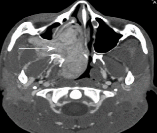
2) CT Scan is the important diagnostic test to know the extent and vascularity of tumor. It can be performed along with angiography.
It usually shows:
- Nasopharyngeal mass.
- Widening of pterygopalatine fissure.
- Enlargement of superior orbital fissure.
- Distortion of nasal septum with erosion of the paranasal sinuses.
- a) Morphology of the lesion.
- b) Soft tissue involvement.
- c) Intracranial extension.

Staging of JNA:
Radkowski staging:
Andrew staging:
Fisch staging:
Newer Endoscopic staging system:
Radkowski staging:
| Stage | Description |
| IA | Involvement limited to the nose and or nasopharynx. |
| IB | Extension into one or more sinus. |
| IIA | Minimal extension into the pterygopalatine fossa. |
| IIB | Occupying the entire pterygopalatine fossa with or without erosion of the orbital apex. |
| IIC | Involvement of the infratemporal fossa with or without extension to the cheek or posterior to the Pterygoid plates. |
| IIIA | Erosion of the skull base (the middle cranial fossa/ base of the pterygoids); minimal intracranial extension. |
| IIIB | Erosion of skull base; Extensive intracranial extension with or without cavernous sinus invasion. |
| Stage | Description |
| I | Tumor limited to nasal cavity and nasopharynx. |
| II | Tumor extension into pterygopalatine fossa, maxillary, sphenoid or ethmoid sinuses. |
| IIIA | Extension into orbit or infratemporal fossa without intracranial extension. |
| IIIB | Stage III A with small extradural intracranial (parasellar) involvement. |
| IVA | Large extradural intracranial or intradural extension. |
| IVB | Extension into cavernous sinus, pituitary or optic chiasma. |
| Stage | Description |
| I | Tumor limited to the nasopharyngeal cavity, bone destruction negligible or limited to the sphenopalatine foramen. |
| II | Tumor invading the pterygopalatine fossa or the maxillary, ethmoid or sphenoid sinus with bone destruction. |
| IIIA | Tumor invading the infratemporal fossa or orbital region without intracranial involvement. |
| IIIB | Tumor invading the infratemporal fossa or orbital region with intracranial extradural (parasellar) involvement. |
| IVA | Intracranial intradural tumor without infiltration of the cavernous sinus, pituitary fossa or optic chiasma. |
| IVB | Intracranial intradural tumor with infiltration of the cavernous sinus, pituitary fossa or optic chiasma. |
| Stage | Description |
| 1 | Nasal cavity, medial parapharyngeal fossa. |
| 2 | Paranasal sinuses, lateral parapharyngeal fossa; no residual vascularity. |
| 3 | Skull base erosion, orbit, infratemporal fossa; no residual vascularity. |
| 4 | Skull base erosion, orbit, infratemporal fossa; residual vascularity. |
| 5 | Intracranial extension, residual vascularity; medial extension; lateral extension. |
Treatment:
1. Surgical excision by:
i. Transnasal maxillary approach.
ii. Transpalatal approach (Wilson’s approach).
iii. Lateral Rhinotomy.
iv. Sublabial mid-facial degloving approach.
v. Infratemporal fossa approach.
vi. Transmaxillary approach (Le fort-I).
vii. Maxillary swing.
viii. Endoscopic resection.
ix. Combined approaches.
A. Combined craniofacial approaches.
B. Combined transpalatal and sublabial (Sardana’s approach).
C. Combined transoral and transnasal endoscopic approach (recent concept). Pre-operative embolization of the tumor drastically reduces the intra-operative hemorrhage.
2. Radiation therapy in usually palliative.
We Are Always Ready to Help You.
Book An Appointment

