FOREIGN BODY OESOPHAGUS
Ingested foreign bodies can lodge in:
- Tonsil: Usually sharp fish bones, needles etc.
- The base of tongue: Fish bone or a needles.
- Pyriform fossa: Fish bone, chicken bone, needle or dentures are commonly seen.
- Oesophagus: Coins, piece of meat, chicken bones, denture, safety pin, marble.
Aetiology:
- Age: Children more prone as they play with coins, marbles and accidently ingest them.
- Loss of protective mechanism: Use of upper denture prevents tactile sensation and a foreign body is swallowed undetected.
- Inadequate mastication.
- Oesophageal stricture, spasm.
- Psychotics.
Constrictions of Oesophagus:

Sites of lodgment of foreign body in the Oesophagus:
- Just below cricopharyngeal sphincter.
- Flat objects like coins are held up at the sphincter while others are held in the upper oesophagus just below sphincter.
- At the broncho-aortic constriction.
- Sharp or pointed foreign bodies can be impacted anywhere in the oesophagus.
Clinical features:
Symptoms:
- History of choking.
- Discomfort or pain just above the clavicle.
- Dysphagia (difficulty in swallowing).
- Drooling of saliva.
- Respiratory distress, dyspnea, cough and wheezing. These symptoms are due to compression overflow or fistulous communication with the air passages.
- Substernal or epigastric pain.
Signs:
- Tenderness in the lower part of neck on the right or left side of trachea.
- Pooling of saliva in pyriform fossa as seen on indirect laryngoscopy.
- Foreign body may be seen protruding from Oesophageal opening in post-cricoid region.
Investigations:
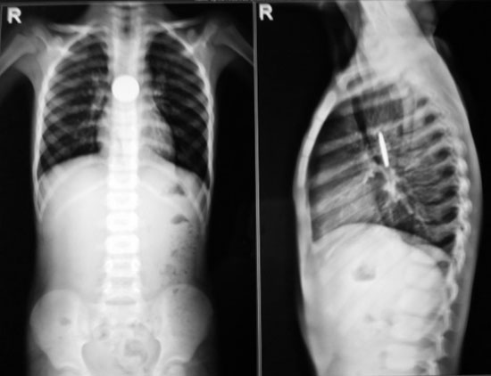
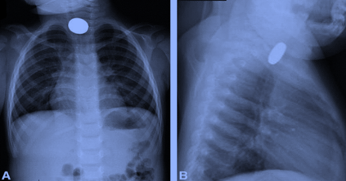
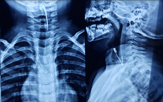
- Plain X-ray can diagnose radio-opaque foreign bodies like coins, safety pin etc. Oesophageal foreign bodies like coins present as a radio-opaque shadow on A-P view while the lateral view shows a vertical slit-like shadow (vice-versa is seen in tracheal foreign bodies).


2. Fluoroscopy to detect hidden foreign bodies and for swallowing function.
3. CT Scan helps to detect small foreign bodies.
Management
- Rigid Oesophagoscopy under general anesthesia is usually safest and the best method of removal of Foreign bodies.
- If such foreign bodies cannot be removed by the above, then transthoracic oesophagotomy is done.
- Blunt non-impacted foreign bodies can be removed by fibre optic oesophagoscopy especially in high risk patients.
Complications:
- Respiratory obstruction.
- Perioesophageal cellulitis.
- Perforation of oesophagus.
- Tracheo-Oesophageal fistula.
Disc batteries:
- Ingestion of disc batteries is a common problem.
- Batteries contain potassium hydroxide, sodium hydroxide and mercury. These contents leak and causes oesophageal injury like stricture, perforation, tracheo-oesophageal fistula, mediastinitis and death.
- Disc battery causes damage to
➣ Mucosa in 1 hr
➣ Muscle coat in 2–4 hr
➣ Perforation of the oesophagus in 8–12 hr
Hence, they should be removed promptly from the oesophagus.
We Are Always Ready to Help You.
Book An Appointment

